


Spinal Evaluation: The Role of Photogrammetry in Postural Assessment
Posted by Renee North, CPEP on 20th Dec 2023
In the domain of assessing spinal health, the advent of photogrammetry has emerged as an effective tool for precisely measuring and analyzing the curvature of the spine. Physicians, both in clinical and research settings, are increasingly recognizing the significance of two-dimensional digital photography as a non-invasive and highly effective method for assessing spinal posture. There are undeniable benefits and applications of photo imaging in postural evaluation, emphasizing its role in improving patient care and contributing valuable insights to research studies.
Understanding Photogrammetry in Spinal Evaluation
Photogrammetry, utilizing two-dimensional digital photography, has become a widely embraced technique for evaluating spinal posture. This method allows orthopedic and neurosurgeons, physical therapists, physiotherapists, sports therapists, other physicians and researchers to conduct a quantitative analysis, capturing subtle changes in posture with precision. Unlike other methods, photogrammetry provides information in both frontal and sagittal planes, offering a comprehensive view of the spine's positioning.
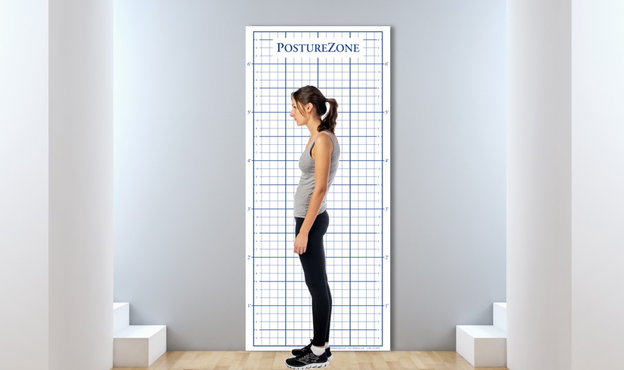
Applications in Clinical Practice
One of the primary advantages of postural photographic images lies in its utility in clinical practice. Physicians can leverage this technique to assess and monitor changes in spinal treatments by comparing quantitative data on posture. By employing standardized wall-mounted grids as reference points, healthcare professionals can enhance the accuracy and reliability of their evaluations, ensuring better-informed decisions for spinal and postural interventions.
Key Concepts in Postural Evaluation
Photographs (posture pictures) proves invaluable in evaluating various aspects of spinal health, including lordosis, kyphosis, and scoliosis. The method allows for the measurement of spinal curvatures, distinguishing between lumbar, thoracic, and cervical curvatures. Through digital analysis, physicians can obtain detailed insights into the angular positioning of the cervical spine, aiding in the assessment of anteriorization of the head.
Standardization and Best Practices
For optimal results, the integration of standardized practices is crucial. Wall Mount grids made from a plastic material, specifically those with a 2" square pattern (approx. 5cm square), have been identified as the preferred reference point for digital postural analysis. Larger than 2 inches makes it more difficult to measure results. Smaller than 2 inches and the image is compromised due to "line-jumping" and blurring of lines in the digital photograph. The use of fixed wall mount posture grids enhances the reliability and durability of the evaluation, especially in clinical, hospital or research settings where consistency is paramount. Banner or vinyl material buckles making precision impossible, and paper grids are not durable and can not be sanitized. It's important to note that portable units, while useful clinically, do not offer the same level of reliability for research studies.
Advantages
Photogrammetry, with its emphasis on standardization and precision, not only contributes to reducing radiation exposure but also facilitates the continuous monitoring of postural treatments. This technique has proven to be an essential tool for clinicians, therapists and researchers, providing a detailed and accurate quantitative evaluation of spinal posture. As the field of spinal evaluation continues to evolve, digital posture evaluation stands as a beacon for best practices, offering a non-invasive, efficient, and reliable approach to understanding and improving spinal and whole body health.